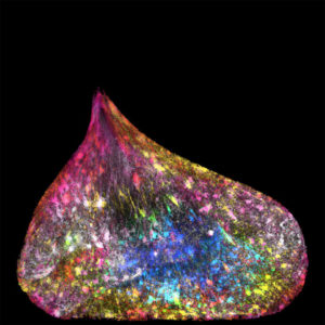
What do you make of the image above? Day Glow slippers under a black light? Colorful sleeping bags for a trio of Minions? March of the Radioactive Penguins?
Of course, it’s none of the above. It’s a very close-up photograph of a developing spinal cord in a mouse. And it’s among the winners of the Wellcome Image Awards 2017, billed as the “most eye-catching celebration of science, medicine and life.”
Eye-catching indeed. The 22 finalists are astonishing, many of them—like the spinal cord at the top of this page—extreme close-ups of cells and tissue from various organisms. And all of them a wonder to behold.
The remarkable imagery moved Dr. Ruth M. Bancewicz to write about how the microscopic, and even the mundane, can move us to look closer and deeper.
Bancewicz counts herself among such scientists. When she read Letters to Malcolm: Chiefly on Prayer, by C.S. Lewis, she was struck by his description of finding “glory” in the little, everyday things.
She writes: “Setting aside intellectual wrestling and doubts, Lewis describes how he allows himself to be inspired by his surroundings: a rippling brook, cushions of moss, or patches of sunlight in a wood. ‘They were not the hope of glory,’ he writes, but ‘they were an exposition of the glory itself. … As it impinges on our will or our understanding, we give it different names—goodness or truth or the like. But its flash upon our sense and mood is pleasure.'”
Take another look at that developing mouse spine. The photographer, Gabriel Galea, is a veterinarian who is studying the development of neural tubes, from which the spinal cord and the central nervous system are formed. Defects in the neural tubes can lead to severe neurodevelopmental disorders, such as spina bifida. Galea hopes his research will reveal why the proper formation of neural tubes fails in around 1 in 1,000 pregnancies.
The picture is a series of three images showing the open end of a mouse’s neural tube. Each image highlights (in blue) one of three main embryonic tissue types—the neural tube itself (left), which develops into the brain, spine, and nerves; the mesoderm (middle), which will form the organs, and the surface ectoderm (right), which will form the skin, teeth and hair.
(Read more details about Galea’s work and a description of his imaging technique—confocal microscopy.)
Bancewicz, who blogs at Science and Belief, concludes her article with these words:
“Life is hectic for a university-based scientist. With teaching, research, supervising students, raising money, managing people, writing, administration, and the new (and very valuable) requirement of explaining your research to the general public, there is very little time to breathe.
“These moments of stepping back to enjoy the beauty of a fantastically detailed image are like shafts of bright sunlight in a week that would be otherwise entirely crazy or stressful. Like a series of gifts dotted throughout a month, year, or career, these experiences are signposts to a greater reality than pressing deadlines might suggest…
“Scientists and non-scientists alike can open a window to let in the fresh air. We can stop, look at the beauties around us, reflect, and wonder.”
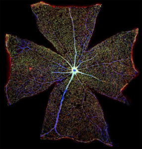
Image credits: Spinal cord by Gabriel Galea, University College London; DNA by Ezequiel Miron, Wellcome Collection; mouse retina by Gabriel Luna, Wellcome Collection.